RNA to Protein : Degradation and Splicing


Molecule is being transported from the nucleus to the cytosol. As its 5ʹ end emerges
from a nuclear pore, the mRNA is met by a ribosome, which begins to translate
it. As translation proceeds, the exon junction complexes (EJCs) that are bound
to the mRNA at each splice site are displaced by the moving ribosome. The normal
stop codon will lie within the last exon, so by the time the ribosome reaches
it and stalls, no more EJCs will be bound to the mRNA. In this case, the mRNA
“passes inspection” and is released to the cytosol where it can be translated in earnest
(Figure 6–76). However, if the ribosome reaches a stop codon earlier, when
EJCs remain bound, the mRNA molecule is rapidly degraded. In this way, the first
round of translation allows the cell to test the fitness of each mRNA molecule as it
exits the nucleus.

Nonsense-mediated decay may have been especially important in evolution,
allowing eukaryotic cells to more easily explore new genes formed by DNA rearrangements,
mutations, or alternative patterns of splicing—by selecting only those
mRNAs for translation that can produce a full-length protein. Nonsense-mediated
decay is also important in cells of the developing immune system, where
the extensive DNA rearrangements that occur.

DNA to RNA : RNA Splicing


Although it may seem at first counterintuitive, the way a gene is packaged into
chromatin can affect how the RNA transcript of that gene is ultimately spliced.
Nucleosomes tend to be positioned over exons (which are, on average, close to the
length of DNA in a nucleosome), and it has been proposed that these act as “speed
bumps,” allowing the proteins responsible for exon definition to assemble on the
RNA as it emerges from the polymerase. In addition, changes in chromatin structure
are used to alter splicing patterns. There are two ways this can happen. First,
because splicing and transcription are coupled, the rate at which RNA polymerase
moves along DNA can affect RNA splicing. For example, if polymerase is moving
slowly, exon skipping (see Figure 6–30A) is minimized: assembly of the initial
spliceosome may be complete before an alternative choice of splice site even
emerges from the RNA polymerase. The nucleosomes in condensed chromatin
can cause polymerase to pause; the pattern of pauses in turn affects the extent of
RNA exposed at any given time to the splicing machinery.


RNA Splicing Shows Remarkable Plasticity

We have seen that the choice of splice sites depends on such features of the premRNA
transcript as the strength of the three signals on the RNA (the 5ʹ and 3ʹ splice
junctions and the branch point) for the splicing machinery, the co-transcriptional
assembly of the spliceosome, chromatin structure, and the “bookkeeping” that
underlies exon definition. We do not know exactly how accurate splicing normally
is because, as we see later, there are several quality control systems that rapidly
destroy mRNAs whose splicing goes awry. However, we do know that, compared
with other steps in gene expression, splicing is unusually flexible.


DNA to RNA : Transcription and Translation


Schematic illustration of an export-ready mRNA molecule and its transport through the nuclear pore. As indicated, some proteins travel with the mRNA as it moves through the pore, whereas others remain in the nucleus. The nuclear export receptor for mRNAs is a complex of proteins that binds to an mRNA molecule once it has been correctly spliced  and polyadenylated. After the mRNA has
been exported to the cytosol, this export receptor dissociates from the mRNA and is re-imported into the nucleus, where it can be used again. The final check indicated here, called nonsense-mediated decay, will be described later in the chapter.


DNA Replication : The Regulated Sliding Clamp


Schematic illustration showing how the clamp (with red and yellow subunits) is loaded onto DNA to serve as a tether for a moving DNA polymerase molecule. The structure of the clamp loader (dark green) resembles a screw nut, with its threads matching the grooves of double-stranded DNA. The loader binds to a free clamp molecule, forcing a gap in its ring of subunits so that this ring is able to slip around DNA. The clamp loader, thanks to its screw-nut structure, recognises the region of DNA that is double-stranded and latches onto it, tightening around the complex of a template strand with a freshly synthesized elongating (primer) strand. It carries the clamp along this double-stranded region until it encounters the 3ʹ end of the primer, at which point the loader hydrolyzes ATP and releases the clamp, allowing it to close around the DNA and bind to DNA polymerase. In the simplified reaction shown here, the clamp loader dissociates into solution once the clamp has been assembled. At a true replication fork, the clamp loader remains close to the polymerase so that, on the lagging strand, it is ready to assemble a new clamp at the start of each new Okazaki fragment


The two proteins shown are present in both bacteria and eukaryotic cells: MutS binds specifically to mismatched base pair, while MutL scans the nearby DNA for a nick. Once Must finds a nick, it triggers the degradation of the nicked strand all the way back through mismatch. Because nicks are largely confined to newly replicated strands in eukaryotes, replication errors are selectively removed. In bacteria, an additional protein in the complex (MutH) nicks unmethylated (and therefore newly replicated) GATC sequences, thereby beginning the process illustrated here. In eukaryotes, MutL contains a DNA nicking activity that aids in the removal of the damaged strands

DNA Replication : The Mehcanism


DNA polymerase performs the first proofreading step just before a new nucleotide
is covalently added to the growing chain. Our knowledge of this mechanism
comes from studies of several different DNA polymerases, including one produced
by a bacterial virus, T7, that replicates inside E. coli. The correct nucleotide
has a higher affinity for the moving polymerase than does the incorrect nucleotide,
because the correct pairing is more energetically favorable. Moreover, after
nucleotide binding, but before the nucleotide is covalently added to the growing
chain, the enzyme must undergo a conformational change in which its “grip”
tightens around the active site (see Figure 5–4). Because this change occurs more
readily with correct than incorrect base-pairing, it allows the polymerase to “double-
check” the exact base-pair geometry before it catalyzes the addition of the
nucleotide. Incorrectly paired nucleotides are harder to add and therefore more
likely to diffuse away before the polymerase can mistakenly add them.
The next error-correcting reaction, known as exonucleolytic proofreading,
takes place immediately after those rare instances in which an incorrect nucleotide
is covalently added to the growing chain. DNA polymerase enzymes are
highly discriminating in the types of DNA chains they will elongate: they require
a previously formed, base-paired 3ʹ-OH end of a primer strand (see Figure 5–4).

Those DNA molecules with a mismatched (improperly base-paired) nucleotide
at the 3ʹ-OH end of the primer strand are not effective as templates because the
polymerase has difficulty extending such a strand. DNA polymerase molecules
correct such a mismatched primer strand by means of a separate catalytic site
(either in a separate subunit or in a separate domain of the polymerase molecule,
depending on the polymerase). This 3ʹ-to-5ʹ proofreading exonuclease clips off any
unpaired or mispaired residues at the primer terminus, continuing until enough
nucleotides have been removed to regenerate a correctly base-paired 3ʹ-OH terminus
that can prime DNA synthesis. In this way, DNA polymerase functions as a
“self-correcting” enzyme that removes its own polymerization errors as it moves
along the DNA (Figure 5–8 and Figure 5–9).

The self-correcting properties of the DNA polymerase depend on its requirement
for a perfectly base-paired primer terminus, and it is apparently not possible
for such an enzyme to start synthesis de novo, without an existing primer.
By contrast, the RNA polymerase enzymes involved in gene transcription do not
need such an efficient exonucleolytic proofreading mechanism: errors in making
RNA are not passed on to the next generation, and the occasional defective RNA
molecule that is produced has no long-term significance. RNA polymerases are
thus able to start new polynucleotide chains without a primer.


DNA Replication : The Introduction


DNA templating is the mechanism the cell uses to copy the nucleotide sequence of one DNA strand into a complementary DNA sequence This process requires the separation of the DNA helix into two template strands, and entails the recognition of each nucleotide in the DNA template strands by a free (unpolymerized) complementary nucleotide.


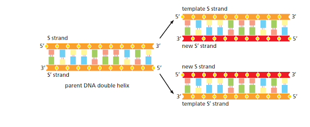
During DNA replication inside a cell, each of the two original DNA strands serves as a template for the formation of an entire new strand. Because each of the two daughters of a dividing cell inherits a new DNA double helix containing one original and one new strand (Figure 5–5), the DNA double helix is said to be replicated “semiconservatively.” How is this feat accomplished?


In all cells, DNA sequences are maintained and replicated with high fidelity. The mutation rate, approximately one nucleotide change per 1010 nucleotides each time the DNA is replicated, is roughly the same for organisms as different as bacteria and humans. Because of this remarkable accuracy, the sequence of the human genome (approximately 3.2 × 10⁹ nucleotide pairs) is unchanged or changed by only a few nucleotides each time a typical human cell divides. This allows most humans to pass accurate genetic instructions from one generation to the next, and also to avoid the changes in somatic cells that lead to cancer.

Centromere : A model for the structure of a Simple


Nucleosomes carrying histone variants have a distinctive character and are thought to be able to produce marks in chromatin that are unusually long-lasting. An important example is seen in the formation and inheritance of the specialized chromatin structure at the centromere, the region of each chromosome required for attachment to the mitotic spindle and orderly segregation of the duplicated copies of the genome into daughter cells each time a cell divides. In many complex organisms, including humans, each centromere is embedded in a stretch of special centromeric chromatin that persists throughout interphase, even though the centromere-mediated attachment to the spindle and movement of DNA occur only during mitosis. This chromatin contains a centromere-specific variant H3 histone, known as CENP-A, plus additional proteins that pack the nucleosomes into particularly dense arrangements and form the kinetochore, the special structure required for attachment of the mitotic spindle.

A specific DNA sequence of approximately 125 nucleotide pairs is sufficient to serve as a centromere in the yeast S. cerevisiae. Despite its small size, more than a dozen different proteins assemble on this DNA sequence; the proteins include the CENP-A histone H3 variant, which, along with the three other core histones, forms a centromere-specific nucleosome. The additional proteins at the yeast centromere attach this nucleosome to a single microtubule from the yeast mitotic spindle.


The Chromatin in Centromeres Reveals How Histone Variants Can Create Special Structures


The centromeres in more complex organisms are considerably larger than those in budding yeasts. For example, fly and human centromeres extend over hundreds of thousands of nucleotide pairs and, while they contain CENP-A, they do not seem to contain a centromere-specific DNA sequence. These centromeres largely consist of short, repeated DNA sequences, known as alpha satellite DNA in humans.

Chromosomal : Packaged into a Set of Chromosomes


Each chromosome in a eukaryotic cell consists of a single, enormously long linear DNA molecule along with the proteins that fold and pack the fine DNA thread into a more compact structure. In addition to the proteins involved in packaging, chromosomes are also associated with many other proteins (as well as numerous RNA molecules). These are required for the processes of gene expression, DNA replication, and DNA repair. The complex of DNA and tightly bound protein is called chromatin (from the Greek chroma, “color,” because of its staining properties).

Bacteria lack a special nuclear compartment, and they generally carry their genes on a single DNA molecule, which is often circular (see Figure 1–24). This DNA is also associated with proteins that package and condense it, but they are different from the proteins that perform these functions in eukaryotes. Although the bacterial DNA with its attendant proteins is often called the bacterial “chromosome, it does not have the same structure as eukaryotic chromosomes, and less is known about how the bacterial DNA is packaged. Therefore, our discussion of chromosome structure will focus almost entirely on eukaryotic chromosomes.

Nucleosomes : Basic Unit of Eukaryotic Chromosome Structure


The proteins that bind to the DNA to form eukaryotic chromosomes are traditionally divided into two classes: the histones and the non-histone chromosomal proteins, each contributing about the same mass to a chromosome as the DNA. The complex of both classes of protein with the nuclear DNA of eukaryotic cells is known as chromatin


Nucleosomes as seen in the electron microscope

(A) Chromatin
isolated directly from an interphase nucleus appears in the electron microscope as a thread about 30 nm thick

(B) This electron micrograph shows a length of chromatin that has been experimentally unpacked, or decondensed, after isolation to show the nucleosomes.

DNA : The Structure and Function


A deoxyribonucleic acid (DNA) molecule consists of two long polynucleotide
chains composed of four types of nucleotide subunits. Each of these chains is
known as a DNA chain, or a DNA strand. The chains run antiparallel to each other,
and hydrogen bonds between the base portions of the nucleotides hold the two
chains together

DOUBLE HELIX

The three-dimensional structure of DNA—the DNA double helix—arises from
the chemical and structural features of its two polynucleotide chains. Because
these two chains are held together by hydrogen-bonding between the bases on
the different strands, all the bases are on the inside of the double helix, and the
sugar-phosphate backbones are on the outside In each case, a
bulkier two-ring base is paired with a single-
ring base (a pyrimidine): A always pairs with T, and G with C


luvne.com ayeey.com cicicookies.com mbepp.com tipscantiknya.com kumpulanrumusnya.comnya.com